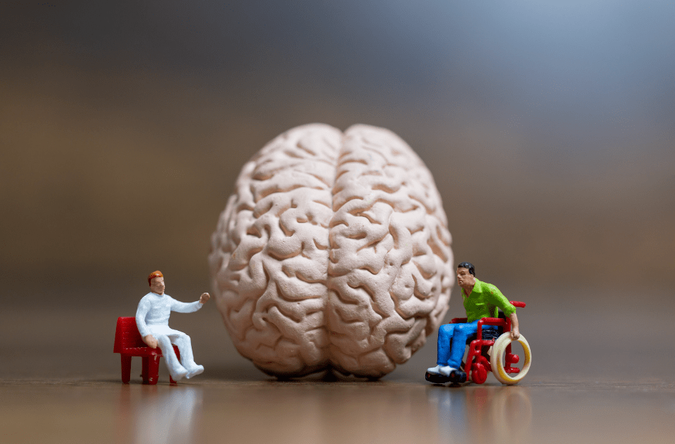
Environmental Enrichment to Promote Neuroplasticity and Prevent Cognitive Decline After Acquired Brain Injury

*Disclaimer: the information on this site is intended as general information only and does not serve as personal medical or therapeutic advice. Use with caution and consult a medical professional familiar with your case beforehand.

**Please note that this blog post is an adaptation of a presentation I created for NeuroRestorative.

The treatment of brain injuries has come a long way in the past 20-30 years and yet even today’s science remains limited when it comes to reversing and preventing the signs and symptoms we see post- moderate to severe brain injury. As a rehabilitation professional I often face questions from the care partners, family members and friends of persons living with brain injuries regarding ways they can help support the health and recovery of that person. Often I am hearing these questions in the first days, weeks, and months post-injury, but this concept is just as important in the years following a brain-injury because while we traditionally think of brain injuries as a one-time injury that either stays the same or gets better over time, more recent research suggests that chronic brain injury is actually a progressive condition. 

As rehab professionals working with neurologic conditions we spend a great deal of time learning about evidence-based treatment techniques for minimizing spasticity, improving gait, maximizing outcomes and so forth. We do our best to engage the patient’s support system in helping them perform a home exercise program and making sure they get to their appointments consistently but what if there was more we could recommend? What if there were changes and modifications that could be made in the living environment, whether that be a house, inpatient rehabilitation facility or long-term care facility, that could support their recovery and prevent later cognitive and functional decline for persons with brain injuries? Turns out…there is!

WAIT, BRAIN INJURIES ARE PROGRESSIVE?

Like most of the conditions we treat, no two brain injuries are alike. Brain injuries can be traumatic or nontraumatic in origin. They can be focal (localized) or diffuse (spread out). They can be caused by bleeding in the brain, loss of oxygen to brain tissue, a tumor compressing brain tissue, infection and more. Their effects are dependent on the location of the injury and available medical treatments to ameliorate the damage to the nerves in the brain. While we would expect that the impairments associated with a brain injury would be progressive in the case of a growing tumor or a spreading infection, we may not think about the progression of symptoms following a stroke or traumatic brain injury where the damage is usually contained to the area of initial injury. 

Within the last ten years, however, scientific research is suggesting that a traumatic brain injury is actually just the starting point of an ongoing physiologic process and is increasingly being understood as a progressive disorder. Imaging studies over time reveal reductions in brain volume and white matter integrity beyond what would be expected to occur after scarring and edema reduction are complete. This subacute deterioration may be linked to deterioration in functional and behavioral outcomes in the years after the initial injury. Individuals who have experienced a moderate to severe TBI also have a higher incidence of developing neurodegenerative conditions like Parkinson’s Disease and Alzheimer’s. 

So what we glean from this information is that while our initial efforts may be placed on recovery and rehabilitation, we cannot forget that interventions aimed at slowing or minimizing this subacute deterioration should also be considered. Since there is a good chance an individual will not be undergoing continuous rehabilitation after their injury, it is important that we educate caregivers and care partners on steps they can take to combat this progression. This brings us to one area of their care that can be modified–the environment.

Tomaszczyk et al., 2014 note that: 

Outside of an intensive rehabilitation program, persons with TBI experience  increased idle time, boredom, and little-to-no engagement in meaningful activities. (Turner et al. 2009; Turner et al. 2007; Frasca et al. 2013). They found that persons with TBI are often isolated from former professional and social networks,  often less socially engaged, feel socially isolated and lonely, and may not be able to resume their previously challenging work activities. (Bulinski 2010; Morton and Wehman 1995). Similarly, physical barriers (e.g., accessibility) and poor communication skills serve to compound already reduced community and social integration (Fleming et al. 2013; Struchen et al. 2011). 

With reduction in leisure activities after a TBI, we see an associated increases in television watching

Reduced schedules of activity in moderate to severe TBI patients have been shown to be associated with poorer neural outcomes. 

Since the schedule of activity and surrounding environment is largely under voluntary control of parents, family, care partners, caregivers, rehab staff, etc…this is one thing we can modify, even into the chronic stages of injury, not just immediately after, to help ensure better long term outcomes for these individuals. 

WHAT IS ENVIRONMENTAL ENRICHMENT?

In the research paradigm, environmental enrichment (EE) refers to a multifaceted form of housing that provides enhanced motor, cognitive, sensory and social stimulation. In animal studies this often includes a living environment with a variety of tunnels, toys and nesting materials that are changed regularly to maintain novelty versus a standard environment with just a couple of things that never change. The theory behind EE is that brains in richer, more stimulating environments have higher rates of synaptogenesis. Animal studies like the ones below provide evidence that EE positively effects outcomes after acquired brain injuries in rats:

So, we know that animals are benefiting from an enriched environment, but how does this relate to brain injuries in humans?

While we usually think about neuroplasticity in a positive light–this is how we promote recovery and compensation–it can also be negative. Several researchers have concluded that negative neuroplastic changes secondary to disuse contributes to chronic cognitive and neural decline. Because a person with a brain injury may be unable or not have ample opportunities to participate in activities that are as stimulating as you and I engage in, neuroplasticity begins working against them.

In the brain, the hippocampus is the center for learning and memory and imaging studies are showing atrophy in this area is correlated to the number of hours of EE a person experiences in the first year post-TBI and that engagement in the simple routines that many of these patients may adopt post-injury are not challenging enough to prevent that hippocampal volume loss. 

DOES THIS WORK FOR HUMANS?

While there is pretty robust evidence for EE in animal studies, the human research studies are ongoing and more limited. I will present a few research studies below that outline some different EE paradigms created for human research but I want to start off by acknowledging the limitations of some of these studies. In order to determine that a particular intervention consistently produces a certain outcome, several things need to occur across multiple research studies. In EE studies, we need to see larger but fairly homogenous groups of participants post-TBI receiving similar types and dosing of EE across multiple settings and different time frames. What you will see below and which was highlighted in the Cochrane Review published in 2021 by Qin et al., is that the heterogeneity between studies at this point does not allow for conclusive recommendations for the application of EE in a population of brain-injured clients. Does this mean that EE is not helpful and we should not implement any of these strategies? No, not at this point. To my knowledge there is not evidence that these interventions are harmful or that they do not work, but we have not had enough of the right types of studies to prove with statistics exactly which aspects of EE are critical for enhancing brain plasticity and what dosing is optimal. 

CONSIDERATIONS FOR IMPLEMENTATION OF EE

One of the things I liked the most about the Jantz (2020) and Kumar & Galloway (2021) studies is that they highlight how these interventions can be applied by non-rehab professionals. While installing an overhead track system (Kumar & Galloway, 2021) may not be feasible for all homes, it underscored how creating opportunities for these adults to participate in self-selected standing activities within their home positively impacted several other mobility and social outcomes. 

Jantz (2020) also showed how goals and impairments identified by a rehab professional were used to create enrichment activities that could be implemented by non-rehab staff and family members. In this scenario the school children got to perform novel and salient activities which actually resulted in better long-term outcomes than just performing pencil push-ups, for example, over and over. 

So how can we take the somewhat limited research and apply it with our clientele, residents, patients and family members? Here are some tips:

As you can see, implementing EE strategies will take some time and should be an effort by all those involved in the person’s care, not just rehab professionals. Taking the time to find out what activities are salient to the target clients is essential and being flexible and responsive to how these are working on a daily basis is important. 

DON’T FORGET TO MONITOR RESPONSE TO ENVIRONMENTAL ENRICHMENT ACTIVITIES

If you are excited about implementing some of these strategies at your facility or to begin educating family members and caregivers of those with brain injuries about them, here are some things to take into consideration. As we talked about in the beginning, every brain injury is different and when it comes to moderate to severe brain injuries, tolerance to new, novel and intense activities can vary from person to person and from day to day. These are some things to evaluate when determining which activities to select and when it may be time to take a break:

  • Fatigue
  • Overstimulation
  • Understimulation
  • Behavioral
  • Cognitive and Motor impairments
  • Vision impairments
  • Hearing Impairments 

WHAT ABOUT BARRIERS TO IMPLEMENTATION?

As with all interventions, it is important that we acknowledge the potential barriers to implementation as it will allow us to problem-solve and prepare. Some common barriers to implementing EE are as follows:

  • Staffing or availability of persons to help
  • Budgeting
  • Space limitations
  • Meeting the needs of many individuals
  • Physical and cognitive barriers to participation
  • Lack of awareness or training of staff and family members/care partners
  • Access to transportation and equipment
  • Lack of specifics around dosing and which EE strategies are best
  • Family support 

If you’ve managed to make it this far, I hope this information gets the wheels turning about how we can further our reach and effectiveness as rehab professionals and care partners for persons with brain injuries. I am excited to see how this area of research continues to evolve over time and whether we can narrow down the dosing and interventions that will be most effective for limiting the progression of a chronic acquired brain injury. 

Resources

Belchev, Z., Boulos, M. E., Rybkina, J., Johns, K., Jeffay, E., Colella, B., Ozubko, J., Bray, M. J. C., di Genova, N., Levi, A., Changoor, A., Worthington, T., Gilboa, A., & Green, R. (2021). Remotely delivered environmental enrichment intervention for traumatic brain injury: Study protocol for a randomised controlled trial. BMJ Open, 11(2), e039767. https://doi.org/10.1136/bmjopen-2020-039767

Bondi, C. O., Klitsch, K. C., Leary, J. B., & Kline, A. E. (2014). Environmental Enrichment as a Viable Neurorehabilitation Strategy for Experimental Traumatic Brain Injury. Journal of Neurotrauma, 31(10), 873–888. https://doi.org/10.1089/neu.2014.3328

Bramlett, H. M., & Dietrich, W. D. (2015). Long-Term Consequences of Traumatic Brain Injury: Current Status of Potential Mechanisms of Injury and Neurological Outcomes. Journal of Neurotrauma, 32(23), 1834–1848. https://doi.org/10.1089/neu.2014.3352

Briones, T. L., Woods, J., & Rogozinska, M. (2013). Decreased neuroinflammation and increased brain energy homeostasis following environmental enrichment after mild traumatic brain injury is associated with improvement in cognitive function. Acta Neuropathologica Communications, 1(1). https://doi.org/10.1186/2051-5960-1-57

Centers for Disease Control and Prevention. (2022, April 5). Stroke facts. Centers for Disease Control and Prevention. Retrieved April 20, 2022, from https://www.cdc.gov/stroke/facts.htm

Centers for Disease Control and Prevention. (n.d.). Traumatic Brain Injury and Concussion: TBI Data. Centers for Disease Control and Prevention (CDC). Retrieved April 25, 2022, from https://www.cdc.gov/traumaticbraininjury/data/

Evans, J. J., Bateman, A., Turner, G., & Green, R. (2008). Research digest. Neuropsychological Rehabilitation, 18(3), 372–384. https://doi.org/10.1080/09602010801909153

Green, R. E. A., Colella, B., Maller, J. J., Bayley, M., Glazer, J., & Mikulis, D. J. (2014). Scale and pattern of atrophy in the chronic stages of moderate-severe TBI. Frontiers in Human Neuroscience, 8. https://doi.org/10.3389/fnhum.2014.00067

International Classification of Functioning, Disability and Health (ICF). (2022, April 29). World Health Organization (WHO). Retrieved April 29, 2022, from https://icd.who.int/dev11/l-icf/en

Jantz, P. B. (2020). Implementing environmental enrichment strategies to help children who have sustained a moderate or severe traumatic brain injury. Support for Learning, 35(3), 276–297. https://doi.org/10.1111/1467-9604.12310

Khan, F., Amatya, B., Elmalik, A., Lowe, M., Ng, L., Reid, I., & Galea, M. (2016). An enriched environmental programme during inpatient neuro-rehabilitation: A randomized controlled trial. Journal of Rehabilitation Medicine, 48(5), 417–425. https://doi.org/10.2340/16501977-2081

Kumar, D. S., & Galloway, J. C. (2021). Feasibility of a home-based environmental enrichment paradigm to enhance purposeful activities in adults with traumatic brain injury: a case series. Disability and Rehabilitation, 1–7. https://doi.org/10.1080/09638288.2020.1868583

Lindberg, R. H. (2021). Nontraumatic brain injury. Brain Injury Medicine, 332–336.e2. https://doi.org/10.1016/b978-0-323-65385-5.00062-7

Masel, B. E., & DeWitt, D. S. (2010). Traumatic Brain Injury: A Disease Process, Not an Event. Journal of Neurotrauma, 27(8), 1529–1540. https://doi.org/10.1089/neu.2010.1358

Matter, A. M., Folweiler, K. A., Curatolo, L. M., & Kline, A. E. (2011). Temporal Effects of Environmental Enrichment–Mediated Functional Improvement After Experimental Traumatic Brain Injury in Rats. Neurorehabilitation and Neural Repair, 25(6), 558–564. https://doi.org/10.1177/1545968310397206

McDonald, M. W., Hayward, K. S., Rosbergen, I. C. M., Jeffers, M. S., & Corbett, D. (2018). Is Environmental Enrichment Ready for Clinical Application in Human Post-stroke Rehabilitation? Frontiers in Behavioral Neuroscience, 12. https://doi.org/10.3389/fnbeh.2018.00135

Miller, L. S., Colella, B., Mikulis, D., Maller, J., & Green, R. E. A. (2013). Environmental enrichment may protect against hippocampal atrophy in the chronic stages of traumatic brain injury. Frontiers in Human Neuroscience, 7. https://doi.org/10.3389/fnhum.2013.00506

Ng, L., Reid, I., Gorelik, A., Galea, M., & Khan, F. (2015). Environmental enrichment for stroke and other non-progressive brain injury. Cochrane Database of Systematic Reviews. https://doi.org/10.1002/14651858.cd011879

Ng, S. Y., & Lee, A. Y. W. (2019). Traumatic Brain Injuries: Pathophysiology and Potential Therapeutic Targets. Frontiers in Cellular Neuroscience, 13. https://doi.org/10.3389/fncel.2019.00528

Quick brain tumor facts. National Brain Tumor Society. (2022, February 1). Retrieved May 1, 2022, from https://braintumor.org/brain-tumor-information/brain-tumor-facts/#additional-info

Scottish Acquired Brain Injury Network (SABIN). (2017). Non-Traumatic Brain Injury – Scottish Acquired Brain Injury Network – e-learning. Scottish Acquired Brain Injury Network – e-Learning. Retrieved April 27, 2022, from https://www.acquiredbraininjury-education.scot.nhs.uk/what-is-acquired-brain-injury/non-traumatic-brain-injury/

Qin, H., Reid, I., Gorelik, A., & Ng, L. (2021). Environmental enrichment for stroke and other non-progressive brain injury. Cochrane Database of Systematic Reviews, 2021(11). https://doi.org/10.1002/14651858.cd011879.pub2

Shors, T., Anderson, M., Curlik, D., & Nokia, M. (2012). Use it or lose it: How neurogenesis keeps the brain fit for learning. Behavioural Brain Research, 227(2), 450–458. https://doi.org/10.1016/j.bbr.2011.04.023

Tomaszczyk, J. C., Green, N. L., Frasca, D., Colella, B., Turner, G. R., Christensen, B. K., & Green, R. E. A. (2014). Negative Neuroplasticity in Chronic Traumatic Brain Injury and Implications for Neurorehabilitation. Neuropsychology Review, 24(4). https://doi.org/10.1007/s11065-014-9273-6

Vora, N. M., Holman, R. C., Mehal, J. M., Steiner, C. A., Blanton, J., & Sejvar, J. (2014). Burden of encephalitis-associated hospitalizations in the United States, 1998–2010. Neurology, 82(5), 443–451. https://doi.org/10.1212/wnl.0000000000000086

World Health Organization. (n.d.). International Classification of Functioning, Disability and Health (ICF). World Health Organization (WHO). Retrieved April 26, 2022, from https://www.who.int/standards/classifications/international-classification-of-functioning-disability-and-health

Multiple Sclerosis, Your Bladder and Physical Therapy?

Guest Post By Dr. Ashley Rawlins, Physical Therapist

@womenshealthbyashley

Inspire Pelvic Health

Let’s talk about urinary incontinence! I know, you may not want to talk about it, but I bet you are curious to learn about it. Urinary incontinence (UI), or bladder leakage, is all too common in those with Multiple Sclerosis (MS). There are areas in the brain, brain stem and spinal cord which work together to control urinary function, so if MS affects any of these specific areas, then communication regarding urination is disrupted, and urinary dysfunction results. Urinary dysfunction is seen in up to 90% of persons with MS1, and 80-100% of those with urinary dysfunction, report UI symptoms2. It’s common, it’s frustrating, it significantly impacts your quality of life3, and if experiencing any balance or vision difficulties, then rushing to the bathroom to prevent leakage may increase your risk for fall related injuries4. I often hear that patients feel this is a difficult topic to discuss with their healthcare providers, so I wanted to bring it up for you in order to demystify, destigmatize, clarify and bring a bit of confidence to seek out the help you need and deserve. 

Did you know that there are physical therapists (PT) out there who specialize in the treatment of UI? That’s where I come in. I am a pelvic health physical therapist, and I specialize in the treatment of neuromuscular and musculoskeletal dysfunction in the abdominal and pelvic areas. This includes bladder dysfunction.

In the normally functioning urinary system, urine is made and emptied into the bladder where it is comfortably stored without leaking until it is full and convenient to empty. Bladder emptying should be easy, pain-free and for the most part, complete. You should be able to comfortably hold urine, and void once every 3-5 hours during the day, and up to one time at night. Urinary function is controlled by a part of our nervous system called the autonomic nervous system, and is largely an unconscious function. It seems simple enough, but the physiology of micturition (the behind the scene workings of emptying your bladder) is complex. When there is disruption to this process, then “urinary dysfunction” can occur. 

Here is a simplified version of what should occur without interruption during normal urinary function:

  • Blood circulates through your kidneys, and the product of this normal filtration process is urine.
  • The urine is then emptied from the kidneys into the bladder, a hollow organ that sits behind the pubic bone, via the ureters.
  • The bladder happily fills with urine without us even sensing it, but at a certain threshold, usually around 200 mL of urine, neurologic signals are sent up the spinal cord to a control center in the brain stem, the Pontine Micturition Center (PMC), and you start to feel like maybe you could go to the bathroom. This “first sensation” is usually easily ignored, and the brain tells the bladder to keep quiet, keep filling for now, and sends a signal back down the spinal cord to do so.
  • Once your bladder has filled to about 400 mL, it becomes a lot harder to ignore. The signal is once again sent up the spinal cord, to the brain stem, and you and your brain agree not to put it off any longer. You make your way to the bathroom, sit down, relax your pelvic floor muscles (more on those late), and then unconsciously the urethral sphincters relax, the muscle around your bladder (detrusor) contracts, and urine is eliminated via the urethra, out of the body.
  • The process continues all day, every day.

Urinary Incontinence and Multiple Sclerosis

When there is a wrench in this system somewhere, UI can occur. Urinary incontinence is the involuntary loss of urine. It is by far the most commonly seen urinary dysfunction in physical therapy. There are three main categories of UI depending on the symptoms you may have: stress urinary incontinence (SUI), urge urinary incontinence (UUI) or mixed urinary incontinence (MUI). Each type of UI may have different contributory factors, aggravating factors and treatment approaches. 

  • Stress urinary incontinence: The involuntary loss of urine because of pressure/physical exertion. For example, urine is lost after sneezing, coughing, laughing, jumping, running, etc. 
  • Urge urinary incontinence: The involuntary loss of urine associated with a strong urge to urinate. This can be a small amount of leakage, or a total loss of the bladder contents5.
  • Mixed urinary incontinence: The involuntary loss of urine associated with stress and/or an urge5.

The causes of SUI can be multifactorial. Common contributing factors include pelvic floor muscle weakness, poor pelvic muscle coordination, urethral deficiencies, detrusor muscle overactivity, poor diet or hydration, constipation, or bad lifestyle habits which make leakage worse. When you have MS, the disease process can complicate these factors. When MS leads to damage of the myelin in the parts of the brain, brain stem, or spinal cord which contribute to urinary function, then the messages regarding urination get disrupted. Your physician is always the first line of defense for examining what medically is contributing to urinary dysfunction, when you have multiple sclerosis. There are many wonderful treatment options including medications, intermittent self-catheterization, nerve stimulation and surgical interventions6. The National MS Society is a good resource for further information on these treatment options, and can be found here. In addition to the neurologic dysfunction that is contributing to bladder symptoms there are behavioral and lifestyle factors, and/or muscle dysfunctions that can worsen bladder symptoms. This is where physical therapy can be really helpful. 

How Physical Therapy Can Help Your Bladder:

There are important muscles and nerves that help your bladder function. With urinary incontinence, we are typically taking a closer look at the function of the muscles and nerves in the abdomen and pelvic floor.

The pelvic floor refers to the group of muscles, nerves and fascia which form a basket at the base of the pelvis. These muscles make up several layers, and run from the pubic bone to the tailbone, left and right between the two sit bones, and encircle and support the bladder, the bowel, the uterus and the vagina in cis females, and the prostate and penis in cis males. When contracted, the pelvic floor muscles draw up and provide a “lift” of your organs towards your head, and they bring the tailbone and pubic bone inwards towards your center, assisting in closure of the pelvic openings (urethra, vagina and rectum), and stability to your pelvic bones. 

Since these muscles are so important in the support and closure of bladder and urethra, then their optimal function is important to prevent urinary leakage. These muscles need to have sufficient strength, length and coordination to help support the complex function of urination. Pelvic physical therapists will work with you to accomplish this. For more information on the pelvic floor, check out this blog.

A pelvic physical therapist will examine the pelvic floor muscles in order to determine what is going on with each patient’s muscles. A good summary of what this examination may entail can be found here. Depending on the findings, physical therapists use a variety of techniques and exercises to decrease the impairments in the muscles and nerves that are affected. Some of the interventions you may see in physical therapy may include:

  • Pelvic floor muscle strengthening exercises: These exercises are more commonly knowns as Kegels but may also include strengthening exercises for muscles in the abdomen, hips, and thighs as imbalance in the muscles in the surrounding areas of the pelvic floor muscles, may also have an adverse effect on urinary incontinence.
  • Neuromuscular Electrical Stimulation (NMES): Electrical stimulation is provided to the pelvic floor muscles, either trans-vaginally or trans-rectally. The exact mechanism of how NMES improves UI is not known, but it is thought that the stimulation either helps to encourage pelvic floor muscle contraction or curbs the detrusor contractions to reduce the overactivity of the bladder7.
  • Electromyography (EMG) Biofeedback: An instrument-based tool that teaches a person how effectively they are using the muscles, using auditory and visual feedback8. In EMG Biofeedback, the muscle activity is measured using external sensors, and displayed for the person to see. This can be helpful in teaching a person how to contract or relax a muscle, depending on what is needed for that individual9.
  • Hydration and diet management: Typically, this consists of optimizing hydration status since drinking too much or too little can make incontinence worse, and diet modification since certain foods can irritate the bladder leading to increased leakage as well.
  • Constipation management
  • Lifestyle strategies: This may include bladder training, techniques to modulate the autonomic nervous system

In persons with MS and urinary incontinence, it is common to see either pelvic floor muscles that are weak and underactive, tight and overactive, muscles that lack coordination, a bladder muscle (detrusor muscles) that is over reactive, or any combination of these10. Physical therapy has been found to provide substantial decreases in urinary leakage, and significant improvements in the quality of life in persons with MS9. Physical therapy interventions such as those described are easy to do, have little to no side effects, and the rewards are significant9. So why not try it!

For more information on how pelvic health physical therapy can help you, check out these websites:

The Academy of Pelvic Health Physical Therapy

Pelvic Rehab

For help finding a qualified pelvic health physical therapist, you can find people in your area here and here.

Resources:

  1. Aharony MS, Lam O, Corcos J. Evaluation of lower urinary tract symptoms in multiple sclerosis patients: Review of the literature and current guidelines. Can Urol Assoc J. 2017; 11(1-2): 61–64. Doi:  10.5489/cuaj.4058
  2. Tubaro A, Puccini F, De Nunzio C, Digesu GA, Elneil S, Gobbi C, Khullar V. Treatment of lower urinary tract symptoms in patient with multiple sclerosis: a systematic review. Curr Urol Rep. 2012; 13(5):335-42. Doi: 10.1007/s11934-012-0266-9 
  3. Litwiller SE, Frohman EM, Zimmern PE. Multiple sclerosis and the urologist. J Urol. 1999; 161(3):743-5.7.
  4. Block V, Rivera M, Melnick M, Allen D. Do physical therapy interventions affect urinary incontinence and quality of life in people with multiple sclerosis. International Journal of Ms Care. 2015; 17(4): 172-80. Doi: 10.7224/1537-2073.2014-031.
  5. Abrams P, Cardozo L, Fall M, Griffiths D, Rosier P, Ulmsten U, Van Kerrevroeck P, Victor A, Wein A. The standardization of terminology in lower urinary tract function: report from the standardization sub-committee of the international continence society. Urology.  61: 37-49. Doi: 10.1016/S0090-4295(02)02243-4.
  6. National Multiple Sclerosis Society. Bladder Dysfunction. National Multiple Sclerosis Society. Accessed 4/10/2021. https://www.nationalmssociety.org/For-Professionals/Clinical-Care/Managing-MS/Symptom-Management/Bladder-Dysfunction
  7. vanBalken MR, Vergunst H, Bemelmans BLH. The use of electrical devices for the treatment of bladder  dysfunction: a review of methods. J Urol. 2004; 172: 846-851.
  8. Lee HJ, Jung KW, Myung SJ. Technique of functional and motility test: how to perform biofeedback for constipation and fecal incontinence. J Neurogastroenterol Motil. 2013; 19: 532-537. 
  9. Alfarra N, Aldosary H, Almefleh S. Do physical therpay interventions improve urinary incontinence and quality of life in patient with multiple sclerosis: A systematic literature review. Phys Med Rehabil Res. 2019. 4:1-6. Doi: 10.15761/PMRR.1000204.
  10. DeRidder D, Vermeulen C, DeSmet F, et al. Clinical assessment of pelvic floor dysfunction in multiple sclerosis. Neruourol Urodyn. 1998;17:337-542.

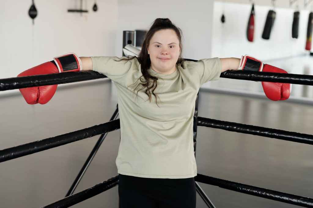
Hittin’ the Gym: Adaptive Exercise Equipment

Photo by Cliff Booth on Pexels.com

It is no surprise that health and fitness remain top priorities for persons with movement impairments. As mobility declines, one’s risk for developing chronic conditions such as heart disease, diabetes or hypertension increases which can add another layer of disability and further impair quality of life. Physical impairments such as difficulty or inability to walk, difficulty using the hands or upper extremities, impairments in balance and so forth can pose a challenge when it comes to going to the gym or participating in an exercise routine. 

The good news is that with a little creativity there are almost always ways to participate in physical exercise that meet your individual needs and abilities.

According to the Physical Activity Guidelines for Amercians 2nd edition, adults with disabilities or chronic conditions should be getting 2.5 to 5 hours of moderate intensity or 75-150 minutes of high intensity activities throughout each week (or a combination of both). Additionally, those that are able to do so should aim to complete at least 2 bouts of strengthening exercises involving major muscles each week (Physical Activity Guidelines for Americans, 2018). In these cases it is important to first get clearance from your doctor that it is safe to begin an exercise plan and then consult with an exercise specialist or physical therapist to attain an individualized exercise program. 

If you are ready to begin exercising many states have adaptive fitness centers outfitted with equipment that might be tailored to your needs. If you are looking to use your local gym facility or stock your own home gym these are examples of equipment that can be used to get you on the path to health and fitness.

**IMPORTANT DISCLAIMER: this information is meant to alert you to the equipment that exists to help you but is not intended as a training module to use it safely. Please contact the manufacturer of each device or a qualified therapist to help you select devices most appropriate to you and to teach you how to use it safely.

**This post may contain affiliate links from Amazon Associates or other affiliate programs through which I may earn a portion of qualifying purchases

Aerobic Conditioning

Recumbent bike/ seated elliptical: a great option for cardiovascular training for those who aren’t confident or able to walk for exercise but can move their legs well enough to use the bike. The Vanswe Recumbent Exercise Bike is a great option for those looking for a reasonable price and good adjustability. The recumbent seat fully supports the back to limit challenges to balance or core strength. Pro tip: if transferring into and out of the seat poses a challenge, look for a model like the HCI Fitness PhysioStep Recumbent Elliptical with Swivel Seat that has a swivel seat that will turn 90 deg to the side to allow for easier access. While certainly pricier, it is likely a better investment than a cheaper model that will end up sitting in the corner collecting dust because it’s too difficult to get in and out of it.

Ski ergometer: this upper body exerciser that can be used in sitting or standing, focuses on building strength in the posterior shoulder and trunk muscle which is important for healthy shoulders and neck especially for wheelchair users. The Concept2 SkiErg is a common model found in some fitness centers but with its narrow design could easily be used in the home too.

Concept2 SkiErg

Vita glide: seated upper body exerciser targets the chest muscles, the shoulder muscles and upper back. The Vita Glide consists of alternating push/pull with the upper extremities and can be used while sitting in a chair.

Upper Body Ergometer (UBE): think of a bicycle for your arms. This device sits on the tabletop and you use your arms to move the pedals forward and back while you sit or stand. These devices range in price and adjustability such as the more basic Vaunn Medical Folding Pedal Exerciser which is fully powered by your arms (or legs) and in comparison the Exerpeutic 2000M Motorized Electric Legs and Arms Pedal Exerciser which actually has a motorized assist program which helps you to move through the full cycle

Exerpeutic 2000M Motorized Electric Legs and Arms Pedal Exerciser

FES bike: FES stands for functional electrical stimulation. While this device comes at a hefty price and is most often seen within rehabilitation clinics some home options are available. This device uses electrical stimulation delivered to the muscles timed with the cycling of the bike to help re-educate muscles through an external stimulus. This device requires training to learn the proper set up and dosing and you should talk with your doctor or therapist to determine if you are a good candidate as any electrical stimulation can carry risks for certain people. The Christopher & Dana Reeve Foundation has a great resource on FES bikes for home use.

Easy Stand Glider: The Easy Stand Glider is a mix between an elliptical and a standing frame. The user can transfer into the seat easily and then through a variety of straps and supports the device will help them into a supported standing position at which point arms and legs can be moved as though standing on an elliptical. This device gives all the benefits of standing with the addition of exercise!

Pedal exercisers: lower extremity exercisers that look like either bike or elliptical pedals. The device can be placed in front of any chair or wheelchair and allow for cardiovascular and leg training. See the links in the Upper Body Ergometer section to see examples of the cycling style and check out this link to see the under desk elliptical style Sunny Health & Fitness Under Desk Elliptical


Wheelchair roller trainer: think of this as a treadmill for a wheelchair. Set up like an indoor trainer for a bicycle, this device allows a manual wheelchair user to simulate forward or backwards motion while remaining stationary. Some models even allow for modification of resistance to allow for greater challenges. These are available for standard manual wheelchair and for some sport or racing chairs. The McLain Wheelchair Trainer is an excellent example of this equipment.

Strengthening

Active Hands: This is an awesome company that creates gripping aids for persons with weak or insufficient grasp or hand strength. The General purpose gripping aid, Looped Exercise aids and Hook aids can be used with most handles, barbells, dumbbells and other strengthening equipment in a commercial or home gym to secure your grasp. 

General Purpose gripping aid

Cuff weights: these weights can take the place of a traditional dumbbell and provide resistance to arms and legs without worrying about having to hold onto the weight. You can buy them in a set of various weights such as the The Deluxe Cuff Ankle and Wrist Weight – 7 Piece Set or individually depending on what you need (BalanceFrom GoFit Fully Adjustable Ankle Wrist Arm Leg Weights)

Medicine ball with strap handles: hands can be inserted into the handles and the straps can be fastened tightly to compensate for weakened grip

FitBALL MedBalls with Straps – 6 lb –

Resistance bands: these bands come in different resistances and provide variable resistance to strengthen muscles throughout the body. Loops can be tied into the ends of the band if grasp is difficult. Bands can be secured to a door or other areas of the home as well to allow for different muscles to be targeted. Bands such as these AZURELIFE Resistance Bands, are a good example.

Rickshaw: The CanDo Rickshaw Rehab Exerciser strengthens arms and shoulders from a seated position. Weight plates can be loaded on the opposite side to increase or decrease resistance. 

CanDo Rickshaw Rehab Exerciser

Other Accessories

Push gloves: often used by individuals with high level spinal cord injuries but also useful for other wheelchair users, push gloves provide additional grip to decrease effort and increase efficiency of pushing the rims of the chair. Useful for everyday use and athletic endeavors. Prime Wheelchair Gloves Protective Gear Mobility Quad-Push Gloves

Elastic straps: For those with weakness in their abdominals or trunk, elastic straps such as these placed around the upper trunk and the back of the chair can better enable the exerciser to lift weights and move their upper body against resistance.

Bodypoint Universal Elastic Strap for Wheelchair

Bibliography

Physical Activity Guidelines for Americans (U.S. Department of Health and Human Services, Compiler; 2nd ed.). (2018). U.S. Department of Health and Human Services. https://health.gov/sites/default/files/2019-09/Physical_Activity_Guidelines_2nd_edition.pdf