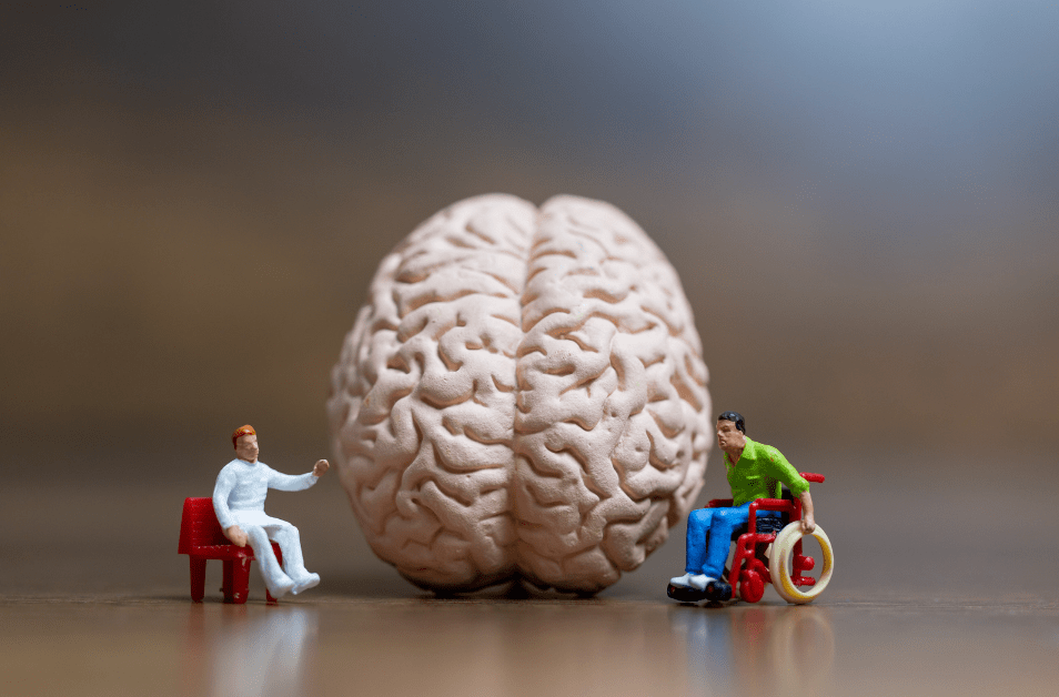
Environmental Enrichment to Promote Neuroplasticity and Prevent Cognitive Decline After Acquired Brain Injury

*Disclaimer: the information on this site is intended as general information only and does not serve as personal medical or therapeutic advice. Use with caution and consult a medical professional familiar with your case beforehand.

**Please note that this blog post is an adaptation of a presentation I created for NeuroRestorative.

The treatment of brain injuries has come a long way in the past 20-30 years and yet even today’s science remains limited when it comes to reversing and preventing the signs and symptoms we see post- moderate to severe brain injury. As a rehabilitation professional I often face questions from the care partners, family members and friends of persons living with brain injuries regarding ways they can help support the health and recovery of that person. Often I am hearing these questions in the first days, weeks, and months post-injury, but this concept is just as important in the years following a brain-injury because while we traditionally think of brain injuries as a one-time injury that either stays the same or gets better over time, more recent research suggests that chronic brain injury is actually a progressive condition. 

As rehab professionals working with neurologic conditions we spend a great deal of time learning about evidence-based treatment techniques for minimizing spasticity, improving gait, maximizing outcomes and so forth. We do our best to engage the patient’s support system in helping them perform a home exercise program and making sure they get to their appointments consistently but what if there was more we could recommend? What if there were changes and modifications that could be made in the living environment, whether that be a house, inpatient rehabilitation facility or long-term care facility, that could support their recovery and prevent later cognitive and functional decline for persons with brain injuries? Turns out…there is!

WAIT, BRAIN INJURIES ARE PROGRESSIVE?

Like most of the conditions we treat, no two brain injuries are alike. Brain injuries can be traumatic or nontraumatic in origin. They can be focal (localized) or diffuse (spread out). They can be caused by bleeding in the brain, loss of oxygen to brain tissue, a tumor compressing brain tissue, infection and more. Their effects are dependent on the location of the injury and available medical treatments to ameliorate the damage to the nerves in the brain. While we would expect that the impairments associated with a brain injury would be progressive in the case of a growing tumor or a spreading infection, we may not think about the progression of symptoms following a stroke or traumatic brain injury where the damage is usually contained to the area of initial injury. 

Within the last ten years, however, scientific research is suggesting that a traumatic brain injury is actually just the starting point of an ongoing physiologic process and is increasingly being understood as a progressive disorder. Imaging studies over time reveal reductions in brain volume and white matter integrity beyond what would be expected to occur after scarring and edema reduction are complete. This subacute deterioration may be linked to deterioration in functional and behavioral outcomes in the years after the initial injury. Individuals who have experienced a moderate to severe TBI also have a higher incidence of developing neurodegenerative conditions like Parkinson’s Disease and Alzheimer’s. 

So what we glean from this information is that while our initial efforts may be placed on recovery and rehabilitation, we cannot forget that interventions aimed at slowing or minimizing this subacute deterioration should also be considered. Since there is a good chance an individual will not be undergoing continuous rehabilitation after their injury, it is important that we educate caregivers and care partners on steps they can take to combat this progression. This brings us to one area of their care that can be modified–the environment.

Tomaszczyk et al., 2014 note that: 

Outside of an intensive rehabilitation program, persons with TBI experience  increased idle time, boredom, and little-to-no engagement in meaningful activities. (Turner et al. 2009; Turner et al. 2007; Frasca et al. 2013). They found that persons with TBI are often isolated from former professional and social networks,  often less socially engaged, feel socially isolated and lonely, and may not be able to resume their previously challenging work activities. (Bulinski 2010; Morton and Wehman 1995). Similarly, physical barriers (e.g., accessibility) and poor communication skills serve to compound already reduced community and social integration (Fleming et al. 2013; Struchen et al. 2011). 

With reduction in leisure activities after a TBI, we see an associated increases in television watching

Reduced schedules of activity in moderate to severe TBI patients have been shown to be associated with poorer neural outcomes. 

Since the schedule of activity and surrounding environment is largely under voluntary control of parents, family, care partners, caregivers, rehab staff, etc…this is one thing we can modify, even into the chronic stages of injury, not just immediately after, to help ensure better long term outcomes for these individuals. 

WHAT IS ENVIRONMENTAL ENRICHMENT?

In the research paradigm, environmental enrichment (EE) refers to a multifaceted form of housing that provides enhanced motor, cognitive, sensory and social stimulation. In animal studies this often includes a living environment with a variety of tunnels, toys and nesting materials that are changed regularly to maintain novelty versus a standard environment with just a couple of things that never change. The theory behind EE is that brains in richer, more stimulating environments have higher rates of synaptogenesis. Animal studies like the ones below provide evidence that EE positively effects outcomes after acquired brain injuries in rats:

So, we know that animals are benefiting from an enriched environment, but how does this relate to brain injuries in humans?

While we usually think about neuroplasticity in a positive light–this is how we promote recovery and compensation–it can also be negative. Several researchers have concluded that negative neuroplastic changes secondary to disuse contributes to chronic cognitive and neural decline. Because a person with a brain injury may be unable or not have ample opportunities to participate in activities that are as stimulating as you and I engage in, neuroplasticity begins working against them.

In the brain, the hippocampus is the center for learning and memory and imaging studies are showing atrophy in this area is correlated to the number of hours of EE a person experiences in the first year post-TBI and that engagement in the simple routines that many of these patients may adopt post-injury are not challenging enough to prevent that hippocampal volume loss. 

DOES THIS WORK FOR HUMANS?

While there is pretty robust evidence for EE in animal studies, the human research studies are ongoing and more limited. I will present a few research studies below that outline some different EE paradigms created for human research but I want to start off by acknowledging the limitations of some of these studies. In order to determine that a particular intervention consistently produces a certain outcome, several things need to occur across multiple research studies. In EE studies, we need to see larger but fairly homogenous groups of participants post-TBI receiving similar types and dosing of EE across multiple settings and different time frames. What you will see below and which was highlighted in the Cochrane Review published in 2021 by Qin et al., is that the heterogeneity between studies at this point does not allow for conclusive recommendations for the application of EE in a population of brain-injured clients. Does this mean that EE is not helpful and we should not implement any of these strategies? No, not at this point. To my knowledge there is not evidence that these interventions are harmful or that they do not work, but we have not had enough of the right types of studies to prove with statistics exactly which aspects of EE are critical for enhancing brain plasticity and what dosing is optimal. 

CONSIDERATIONS FOR IMPLEMENTATION OF EE

One of the things I liked the most about the Jantz (2020) and Kumar & Galloway (2021) studies is that they highlight how these interventions can be applied by non-rehab professionals. While installing an overhead track system (Kumar & Galloway, 2021) may not be feasible for all homes, it underscored how creating opportunities for these adults to participate in self-selected standing activities within their home positively impacted several other mobility and social outcomes. 

Jantz (2020) also showed how goals and impairments identified by a rehab professional were used to create enrichment activities that could be implemented by non-rehab staff and family members. In this scenario the school children got to perform novel and salient activities which actually resulted in better long-term outcomes than just performing pencil push-ups, for example, over and over. 

So how can we take the somewhat limited research and apply it with our clientele, residents, patients and family members? Here are some tips:

As you can see, implementing EE strategies will take some time and should be an effort by all those involved in the person’s care, not just rehab professionals. Taking the time to find out what activities are salient to the target clients is essential and being flexible and responsive to how these are working on a daily basis is important. 

DON’T FORGET TO MONITOR RESPONSE TO ENVIRONMENTAL ENRICHMENT ACTIVITIES

If you are excited about implementing some of these strategies at your facility or to begin educating family members and caregivers of those with brain injuries about them, here are some things to take into consideration. As we talked about in the beginning, every brain injury is different and when it comes to moderate to severe brain injuries, tolerance to new, novel and intense activities can vary from person to person and from day to day. These are some things to evaluate when determining which activities to select and when it may be time to take a break:

  • Fatigue
  • Overstimulation
  • Understimulation
  • Behavioral
  • Cognitive and Motor impairments
  • Vision impairments
  • Hearing Impairments 

WHAT ABOUT BARRIERS TO IMPLEMENTATION?

As with all interventions, it is important that we acknowledge the potential barriers to implementation as it will allow us to problem-solve and prepare. Some common barriers to implementing EE are as follows:

  • Staffing or availability of persons to help
  • Budgeting
  • Space limitations
  • Meeting the needs of many individuals
  • Physical and cognitive barriers to participation
  • Lack of awareness or training of staff and family members/care partners
  • Access to transportation and equipment
  • Lack of specifics around dosing and which EE strategies are best
  • Family support 

If you’ve managed to make it this far, I hope this information gets the wheels turning about how we can further our reach and effectiveness as rehab professionals and care partners for persons with brain injuries. I am excited to see how this area of research continues to evolve over time and whether we can narrow down the dosing and interventions that will be most effective for limiting the progression of a chronic acquired brain injury. 

Resources

Belchev, Z., Boulos, M. E., Rybkina, J., Johns, K., Jeffay, E., Colella, B., Ozubko, J., Bray, M. J. C., di Genova, N., Levi, A., Changoor, A., Worthington, T., Gilboa, A., & Green, R. (2021). Remotely delivered environmental enrichment intervention for traumatic brain injury: Study protocol for a randomised controlled trial. BMJ Open, 11(2), e039767. https://doi.org/10.1136/bmjopen-2020-039767

Bondi, C. O., Klitsch, K. C., Leary, J. B., & Kline, A. E. (2014). Environmental Enrichment as a Viable Neurorehabilitation Strategy for Experimental Traumatic Brain Injury. Journal of Neurotrauma, 31(10), 873–888. https://doi.org/10.1089/neu.2014.3328

Bramlett, H. M., & Dietrich, W. D. (2015). Long-Term Consequences of Traumatic Brain Injury: Current Status of Potential Mechanisms of Injury and Neurological Outcomes. Journal of Neurotrauma, 32(23), 1834–1848. https://doi.org/10.1089/neu.2014.3352

Briones, T. L., Woods, J., & Rogozinska, M. (2013). Decreased neuroinflammation and increased brain energy homeostasis following environmental enrichment after mild traumatic brain injury is associated with improvement in cognitive function. Acta Neuropathologica Communications, 1(1). https://doi.org/10.1186/2051-5960-1-57

Centers for Disease Control and Prevention. (2022, April 5). Stroke facts. Centers for Disease Control and Prevention. Retrieved April 20, 2022, from https://www.cdc.gov/stroke/facts.htm

Centers for Disease Control and Prevention. (n.d.). Traumatic Brain Injury and Concussion: TBI Data. Centers for Disease Control and Prevention (CDC). Retrieved April 25, 2022, from https://www.cdc.gov/traumaticbraininjury/data/

Evans, J. J., Bateman, A., Turner, G., & Green, R. (2008). Research digest. Neuropsychological Rehabilitation, 18(3), 372–384. https://doi.org/10.1080/09602010801909153

Green, R. E. A., Colella, B., Maller, J. J., Bayley, M., Glazer, J., & Mikulis, D. J. (2014). Scale and pattern of atrophy in the chronic stages of moderate-severe TBI. Frontiers in Human Neuroscience, 8. https://doi.org/10.3389/fnhum.2014.00067

International Classification of Functioning, Disability and Health (ICF). (2022, April 29). World Health Organization (WHO). Retrieved April 29, 2022, from https://icd.who.int/dev11/l-icf/en

Jantz, P. B. (2020). Implementing environmental enrichment strategies to help children who have sustained a moderate or severe traumatic brain injury. Support for Learning, 35(3), 276–297. https://doi.org/10.1111/1467-9604.12310

Khan, F., Amatya, B., Elmalik, A., Lowe, M., Ng, L., Reid, I., & Galea, M. (2016). An enriched environmental programme during inpatient neuro-rehabilitation: A randomized controlled trial. Journal of Rehabilitation Medicine, 48(5), 417–425. https://doi.org/10.2340/16501977-2081

Kumar, D. S., & Galloway, J. C. (2021). Feasibility of a home-based environmental enrichment paradigm to enhance purposeful activities in adults with traumatic brain injury: a case series. Disability and Rehabilitation, 1–7. https://doi.org/10.1080/09638288.2020.1868583

Lindberg, R. H. (2021). Nontraumatic brain injury. Brain Injury Medicine, 332–336.e2. https://doi.org/10.1016/b978-0-323-65385-5.00062-7

Masel, B. E., & DeWitt, D. S. (2010). Traumatic Brain Injury: A Disease Process, Not an Event. Journal of Neurotrauma, 27(8), 1529–1540. https://doi.org/10.1089/neu.2010.1358

Matter, A. M., Folweiler, K. A., Curatolo, L. M., & Kline, A. E. (2011). Temporal Effects of Environmental Enrichment–Mediated Functional Improvement After Experimental Traumatic Brain Injury in Rats. Neurorehabilitation and Neural Repair, 25(6), 558–564. https://doi.org/10.1177/1545968310397206

McDonald, M. W., Hayward, K. S., Rosbergen, I. C. M., Jeffers, M. S., & Corbett, D. (2018). Is Environmental Enrichment Ready for Clinical Application in Human Post-stroke Rehabilitation? Frontiers in Behavioral Neuroscience, 12. https://doi.org/10.3389/fnbeh.2018.00135

Miller, L. S., Colella, B., Mikulis, D., Maller, J., & Green, R. E. A. (2013). Environmental enrichment may protect against hippocampal atrophy in the chronic stages of traumatic brain injury. Frontiers in Human Neuroscience, 7. https://doi.org/10.3389/fnhum.2013.00506

Ng, L., Reid, I., Gorelik, A., Galea, M., & Khan, F. (2015). Environmental enrichment for stroke and other non-progressive brain injury. Cochrane Database of Systematic Reviews. https://doi.org/10.1002/14651858.cd011879

Ng, S. Y., & Lee, A. Y. W. (2019). Traumatic Brain Injuries: Pathophysiology and Potential Therapeutic Targets. Frontiers in Cellular Neuroscience, 13. https://doi.org/10.3389/fncel.2019.00528

Quick brain tumor facts. National Brain Tumor Society. (2022, February 1). Retrieved May 1, 2022, from https://braintumor.org/brain-tumor-information/brain-tumor-facts/#additional-info

Scottish Acquired Brain Injury Network (SABIN). (2017). Non-Traumatic Brain Injury – Scottish Acquired Brain Injury Network – e-learning. Scottish Acquired Brain Injury Network – e-Learning. Retrieved April 27, 2022, from https://www.acquiredbraininjury-education.scot.nhs.uk/what-is-acquired-brain-injury/non-traumatic-brain-injury/

Qin, H., Reid, I., Gorelik, A., & Ng, L. (2021). Environmental enrichment for stroke and other non-progressive brain injury. Cochrane Database of Systematic Reviews, 2021(11). https://doi.org/10.1002/14651858.cd011879.pub2

Shors, T., Anderson, M., Curlik, D., & Nokia, M. (2012). Use it or lose it: How neurogenesis keeps the brain fit for learning. Behavioural Brain Research, 227(2), 450–458. https://doi.org/10.1016/j.bbr.2011.04.023

Tomaszczyk, J. C., Green, N. L., Frasca, D., Colella, B., Turner, G. R., Christensen, B. K., & Green, R. E. A. (2014). Negative Neuroplasticity in Chronic Traumatic Brain Injury and Implications for Neurorehabilitation. Neuropsychology Review, 24(4). https://doi.org/10.1007/s11065-014-9273-6

Vora, N. M., Holman, R. C., Mehal, J. M., Steiner, C. A., Blanton, J., & Sejvar, J. (2014). Burden of encephalitis-associated hospitalizations in the United States, 1998–2010. Neurology, 82(5), 443–451. https://doi.org/10.1212/wnl.0000000000000086

World Health Organization. (n.d.). International Classification of Functioning, Disability and Health (ICF). World Health Organization (WHO). Retrieved April 26, 2022, from https://www.who.int/standards/classifications/international-classification-of-functioning-disability-and-health

The Neurologic Screen: what to test, why to test it and when to do it

Photo by Alex Green on Pexels.com

*Disclaimer: the information on this site is intended as general information only and does not serve as personal medical or therapeutic advice. Use with caution and consult a medical professional familiar with your case beforehand.

If you work in pediatric therapy, OP neuro rehab, acute care or inpatient rehabilitation settings you will most likely find yourself needing to do elements of a neuro screen during your evaluations. Even if you decided well before you graduated PT school that you would never work with neurologic patients and you were going to dedicate your career to sports and orthopedic rehab you will undoubtedly encounter scenarios in which you need to do at least some elements of the neurologic screen. If you haven’t ever encountered this, there is a good chance you missed out on an important opportunity to screen for neurologic impairments in at least some of your patients. Unlike a neurologic examination/evaluation that many of us perform on a patient with a known neurologic condition, I consider the neurologic screen to be a tool that helps inform therapists whether the nervous system may be involved in a patient that does not have a known neurologic condition, to help differentially diagnose an impairment that may be undiagnosed or unrecognized or to provide valuable information about nervous system function that could impact the plan of care.

Despite our comprehensive education in physical therapy school, I bet all practicing therapists have been faced with the reality that unless you use a particular area of knowledge or skill pretty regularly, you lose your familiarity and comfort with it pretty quickly. Nothing to be ashamed of, we can’t be experts in everything, but having a basic comfort with the elements of the neuro screen will go a long way in helping therapists create a more comprehensive and effective plan of care and also help identify patients that need to be referred back to their doctors for more testing or medical care. Also, so it doesn’t seem as though I’m just picking on the ortho therapists out there, Neuro Folks, if you aren’t screening your patients for and addressing their orthopedic problems, you’re missing an important piece also!

What’s in a Neuro Screen?

**Alright, quick forewarning here that this is not a completely comprehensive outline of every possible neurologic test you can perform on your patient. You can find that in your neuro rehab textbook.

Performing a good neuro screen requires both an understanding of the purpose of each neurologic test and measure and an ability to determine when to use what test. As a student physical therapist or novice clinician, the clinical reasoning process often involves administering a lot of subjective and objective outcome measures, looking at the results for anything that suggests an impairment or functional limitation then deriving a PT diagnosis and plan of care based on the results of those tests. As clinicians gain experience they often draw very heavily on their understanding of injury and disease and their past patient experiences to make hypotheses that allow them to immediately narrow down the tests and measures they believe will be most pertinent.

If you aren’t as comfortable with the myriad of neurologic screening tools out there and haven’t worked with that population of patients for a long time or very often, use the information below as a jumping off point for selecting tests that should help you recognize how the neurologic and neuromuscular systems are impacted in your patients.

Should I throw the kitchen sink at them?

If you are lucky enough to work in a magical PT clinic where you can spend 2 hours evaluating your patient on the first day then by all means, have at it. If you don’t work in inpatient rehab and you don’t happen to work in a setting that allows for much longer 1:1 sessions with the therapist then you need to be selective and judicious when structuring your exams.

Scenario 1: Patient arrives with a known neurologic diagnosis (Stroke, Multiple Sclerosis, Neuropathy, etc.)

Approach: Use your knowledge about the patient’s injury or condition and the impairments and activity/participation limitations commonly associated with that injury/condition to select your tests/measures. For example, if your patient has neuropathy you would expect that motor and/or sensory systems may be affected so sensation and strength testing would be appropriate. If your patient had a stroke, you might expect changes in muscle tone as well as strength and sensation so you would screen these areas.

Because you do not have time to perform every test in the book, focus on the elements that provide information that will impact your treatment and prognosis. For example, reflex testing certainly provides valuable information about the state of the nervous system following a stroke but if the results don’t change your plan of care then they wouldn’t be my top priority.

Scenario 2: Patient arrives with a known diagnosis that you would not consider to be neurologic in nature but their history or your observation suggests the neurologic system could be contributing to their complaints

Approach: Something in your subjective or objective exam has prompted you to think that this could be neurologic in nature. In this scenario I would start by selecting tests that point to abnormalities in neurologic function such as sensation testing which could point to a dermatomal pattern of sensation loss, for example, coordination testing which can indicate cerebellar involvement, reflex testing which can help distinguish between an upper or lower motor nerve disorder, myotome testing which can indicate spinal nerve involvement or pathologic reflexes like Babinski or Hoffman’s that indicate loss of cortical inhibition and an upper motor nerve lesion.

Scenario 3: Patient arrives with a known neurologic diagnosis but your well-honed spidey-senses suspect they could have been misdiagnosed or there could be a secondary problem happening

Approach: What about this patient’s presentation has led you to believe their is more to this picture than the diagnosis they walked in with? Use this information to select your tests and measures. For example, I once evaluated a patient who had been treated for Guillain Barre Syndrome in the hospital and inpatient rehab and came to my outpatient clinic about 5 months later. On initial evaluation they presented with severe extensor tone and spasticity in the right lower extremity. This gave me pause because GBS is a lower motor neuron disorder while spasticity is an upper motor neuron sign. This finding prompted me to examine the patient further and refer them for more imaging which revealed they had been misdiagnosed and actually had either Transverse Myelitis or Multiple Sclerosis.

In these cases it is a good time to whip out your cranial nerve screens, tone assessments, pathological reflex testing, deep tendon reflex testing, coordination testing and vestibular testing among others to see if you can garner additional information that may indicate the patient should be referred back to their provider for further testing.

Many Tests to Choose From

Now that you have an idea of how to select what body structure/function you want to examine, here is a list of some helpful tests and measures that you can use to inform your plan of care. Please note this list does not include functional outcome measures that assess things like balance, gait or functional strength as I consider those to have a different purpose than a neurologic screen.

Cranial Nerve Screen

Purpose: used to identify lesions of the cranial nerves which can help localize pathology, identify areas that may impact patient response to therapy and recovery or prompt a referral to another provider for treatment.

Sensory Screen:

Purpose: localize lesion, assess protective sensation, monitor the extent of neurologic recovery or disease progression; prognosticate functional outcomes

Basic sensory screen for impairments in light touch, sharp/dull, temperature and vibration can indicate location of lesion:


  • Dermatomal pattern of sensory loss: indicates which spinal levels and tracts are involved
    • Light touch: Ventral Spinothalamic Tract of spinal cord 
    • Pain (pin prick): Lateral Spinothalamic Tract of spinal cord
    • Temperature: Lateral Spinothalamic Tract of spinal cord
    • Proprioception: Dorsal Column of spinal cord
    • Vibration: Dorsal Column of spinal cord
  • Peripheral nerve distribution: indicates an injury or lesion to a particular peripheral sensory nerve
  • Stocking glove pattern: usually consistent with peripheral neuropathy in which nerves are affected based on length of axon and not specific branches of nerves
  • Larger areas affected on one side such as entire leg, arm, face: often points to upper motor nerve lesions involving multiple levels of the spinal cord or the sensory cortex of the brain

Cortical Sensory Testing assesses the function of the sensory association areas of the parietal lobe

  • Stereognosis
  • Graphesthesia
  • Two point discrimination
  • Localization of touch

**Clinical pearl: if you observe a patient walking unsteadily with wide base of support, possibly stomping the ground they may have a sensory ataxia caused by loss of sensation in their feet and legs. Sensation testing should help pick this up and symptoms will worsen significantly in the dark or with eyes closed.

Reflex Testing

Purpose: Deep Tendon Reflexes: assesses muscle stretch reflex and can be localized to specific nerve roots. Hyperreflexia can indicate an upper motor nerve lesion while hypo– or arreflexia can indicate a lower motor nerve lesion

  • Pathologic Reflexes
    • Hoffman’s Reflex: positive Hoffman’s sign (adduction and flexion of the thumb and flexion of the index finger) is suggestive of cortocospinal tract dysfunction in the cervical spinal cord
    • Babinski: positive Babinski (extension of the great toe and splaying of the other toes) is suggestive of a corticospinal tract dysfunction
    • Clonus: rhythmic and oscillating stretch reflex, form of hyperreflexia and indicative of an upper motor neuron lesion

Muscle Tone Assessment

Purpose: Assess the muscle’s response to passive stretch. Lower motor neuron lesions reduce muscle tone while upper motor lesions increase muscle tone

  • Hypertonicity: umbrella term that refers to an increased resistance to passive stretch
    • Includes spasticity, rigidity, dystonia, co-contraction, clonus
  • Spasticity: specifically refers to velocity-dependent resistance to passive stretch
    • Can measure with the Modified Ashworth Scale or the Tardieu 

Coordination Testing

Purpose: usually considered to be a window into cerebellar function so abnormalities in these tests are concerning for cerebellar dysfunction.

    • Rapid Alternating Movement: alternating pronation/supination or ankle plantarflexion/dorsiflexion–the cerebellum controls the ability to perform one movement quickly followed by its opposite. Impairment in this function is called dysdiadochokinesis (if you can spell this word correctly without looking it up, you are my hero).
    • Finger to Nose: measures upper extremity coordination. During this test you may observe
      • Dysmetria: misjudging the target (hypo vs hypermetric movements)
      • Intention tremor 
      • Dyssynergia: refers to breakdown of movement, often see the movement broken in to several smaller movements
      • Ataxia: refers to abnormal coordination of movement
    • Heel to Shin: lower extremity coordination, may be dysmetric (missing target) or generally ataxic

**Clinical pearl: be aware that vision, executive function, cognition, strength and motor control can all play a role in being able to perform these coordination tests well and may confuse the results. Speeding up the movement can unmask coordination impairments

Myotome Testing

Purpose: Weakness is a common complaint in patients receiving physical therapy and may be due to numerous causes. Keep your eye out, however, for patterns of weakness that fall into a myotome pattern as this indicates spinal nerve involvement.

Vestibular and Oculomotor Screen

These tests are most often performed in the context of complaints of dizziness or disequilibrium and in patients with a known neurologic condition. However, if you have a familiarity with some of these tests you may be able to pick up on an issue that may be pertinent to their care and need further evaluation by a specialist. There is a whole litany of tests available to you to differentiate between possible causes of symptoms but these tests may help to at least determine if more in depth assessment is warranted.

Oculomotor Screen: these impairments may manifest as blurry vision, double vision, difficulty reading, impaired eye movements, dizziness, headaches, etc.

    • Smooth pursuit: measures the eyes ability to smoothly track a moving object while the head remains in place. Also allows assessment of strength and function of the ocular muscles. Saccadic intrusions, meaning the movement is broken down into several smaller movements suggests central pathology.
    • Saccades: the ability of the eye to move quickly and accurately from one target to another. Impairments typically suggest cerebellar disorder
    • Convergence: the ability to adduct the eyes and view a near-target object without double vision. Normal convergence is 6-10cm. Doubling of vision before 10cm can be suggestive of a concussion, a cerebellar or brainstem injury or an impairment in eye muscle function

Vestibular Screen: these impairments are often accompanied by dizziness, vertigo, disequilibrium, falls, nausea, disorientation, feeling like you or the world is moving

    1. Vestibular-Ocular Reflex (VOR): the ability to maintain eyes locked on an unmoving target while the head is moving. Use a metronome set at 2Hz and have patient perform oscillations of the head in the sagittal and then transverse plane about 30 deg to each side while locking eyes on a small target about arm’s length away. If they experience symptoms of dizziness, nausea or headache, double vision or blurring then this indicates likely VOR impairment.
    2. Head Impulse Test: failure to maintain eyes locked on therapist’s nose while quickly turning the head to one side suggests peripheral vestibular dysfunction.
    3. VOR cancellation: reflects the ability to suppress VOR and turn eyes and head in same direction at same rate. Normal: eyes maintained in center of orbit while head and eyes track an object moving across the visual field. ABNORMAL: eye cannot track at same rate and will not remain in center of orbit which suggests cerebellar dysfunction.
    4. Nystagmus assessment
      • Positional nystagmus: test with Dix-Hallpike, Roll test or Sidelying Test (Semant Diagnostic Maneuver). NOTE–central causes of positional nystagmus do exist and will not respond to canalith repositioning
      • Spontaneous Nystagmus: occurs from imbalance in vestibular signals being sent to oculomotor nerves. Leads to constant drift of eyes to one direction with brief corrections back the other way. Occurs after acute vestibular loss  
          • Peripheral lesion: fixing gaze on stable object will stop nystagmus, typically resolves within 24 hours.
          • Central: nystagmus persists with fixation on nonmoving object
      • Peripheral Nystagmus characteristics:
        • Direction fixed and dominantly horizontal
        • Suppressed with visual fixation
        • More likely to be exacerbated when looking in direction of fast component
      • Central Nystagmus characteristics
        • Direction changing, more likely to be purely vertical or torsional
        • Not diminished with fixation
        • May be accompanied by one of the “D” symptoms: Diplopia, dysarthria, dysphagia, drop attacks, dyskinesia/ataxia (and muscle weakness on one side).

Put it into Practice

Now that you have a sense of what tests are available and what their results mean, keep an eye out for opportunities to practice them. Continuing education, online modules, textbooks and phone-a-neuro-therapist-friend are all great ways to build your confidence and knowledge in this area.

Dizziness and COVID-19

Photo by CDC on Pexels.com

As the COVID-19 virus and its ever evolving variants continue to infect, at times, hundreds of thousands of people a day worldwide, medical professionals and sufferers of the disease are still working to put together a comprehensive list of typical signs and symptoms. Two clinical manifestations that have arisen and distinguish the COVID-19 virus from many other respiratory viruses are the symptoms of dizziness and vertigo. 

Drs. Saniasiaya and Kulaseharah wrote in the Ear, Nose and Throat Journal in September of 2020 that dizziness has proven to be a “notable clinical manifestation among COVID-19 patients” (Saniasiaya, MD, MMED & Kulasegarah, MB Bch, BAO, MD, 2020). Several causative mechanisms were proposed amongst doctors and researchers but all agreed that when dizziness and vertigo are present, it is important to consider possible causes such as acute labyrinthitis, vestibular neuritis, acute otitis media or even secondary to stroke caused by COVID-19. Each of these potential causes of vertigo and dizziness require their own assessment and treatment by a healthcare professional.

Vestibular neuritis, for example, a condition in which the vestibular nerve that supplies the inner ear organs becomes inflamed, can cause severe (though typically temporary) dizziness and vertigo, feelings of imbalance and nausea. Recovery times can vary from days to months. In a case series published in the Le Infezioni in Medicina journal in March 2021 the authors describe 6 cases in which female patients with known or suspected COVID-19 infections experienced severe dizziness that was later diagnosed as vestibular neuritis (Malayala et al., 2021). The mechanism of this manifestation may be similar to the causes of other symptoms of COVID infection such as loss of smell, changes in vision and changes in the vascular system but can have a lasting impact on some people even once the initial infection seems to have resolved.  While the symptoms may be initially severe, treatment with medications to manage symptoms of nausea and dizziness and decrease inflammation are typical in the acute phase while physical therapy and vestibular rehabilitation can help immensely for any persisting symptoms.

How do I know if I need vestibular rehab?

Vestibular rehabilitation focuses on improving the effectiveness of our vestibular system, the sensory organs in our body that transmit information about how our head is oriented and moving in space. The vestibular system is one of three sensory systems in our body that help us to balance. The second system is the somatosensory system which consists of sensory nerves in the joints and skin, which transmit information such as whether you are standing with your feet flat on the ground or if part of your foot is lifted. The third system is our visual system. Our eyes reference the environment around us to understand better how our body is oriented in space. 

When the vestibular system is affected by an infection such as in vestibular neuritis, an injury like a concussion, or even when small crystals in the inner ear organs get displaced (a condition called BPPV), individuals can become over reliant on the other two sensory systems and sometimes that can persist, “uncompensated” for long periods of time. 

In these cases one might be left with persistent feelings of imbalance, dizziness and disequilibrium. Suddenly closing your eyes in the shower becomes a fight to keep from falling, turning your head to look at traffic while riding in the car makes your head spin and the thought of going on a hike makes you nervous for your safety. Not to worry though, a physical therapist who is experienced in vestibular rehabilitation can assess your symptoms and prescribe exercises to help reduce your residual symptoms and help to restore your function. In many cases the vestibular system can be trained to perform at a higher level again. There is no need to keep living with these symptoms long term!

So if you or someone you know is experiencing severe dizziness along with any of the other known symptoms of COVID (ie. sore throat, fever, cough, etc.), talk to your doctor about what treatment options are available to help reduce your symptoms quickly and to rule out more serious causes of dizziness like a stroke. If you find yourself struggling to get back to normal after COVID and are feeling off balance or dizzy, seek care from an experienced physical therapist and they will help get you back on the road to full recovery! 

Bibliography

Malayala, S. V., Mohan, G., Vasireddy, D., & Atluri, P. (2021). A case series of vestibular symptoms in positive or suspected COVID-19 patients. Le Infezioni in Medicina, 29(1), 117-122. https://www.infezmed.it/index.php/article?Anno=2021&numero=1&ArticoloDaVisualizzare=Vol_29_1_2021_117

Saniasiaya, MD, MMED, J., & Kulasegarah, MB Bch, BAO, MD, J. (2020, September 15). Dizziness and COVID-19. Ear Nose and Throat Journal, 100(1), 29-30. https://journals.sagepub.com/doi/10.1177/0145561320959573

Multiple Sclerosis, Your Bladder and Physical Therapy?

Guest Post By Dr. Ashley Rawlins, Physical Therapist

@womenshealthbyashley

Inspire Pelvic Health

Let’s talk about urinary incontinence! I know, you may not want to talk about it, but I bet you are curious to learn about it. Urinary incontinence (UI), or bladder leakage, is all too common in those with Multiple Sclerosis (MS). There are areas in the brain, brain stem and spinal cord which work together to control urinary function, so if MS affects any of these specific areas, then communication regarding urination is disrupted, and urinary dysfunction results. Urinary dysfunction is seen in up to 90% of persons with MS1, and 80-100% of those with urinary dysfunction, report UI symptoms2. It’s common, it’s frustrating, it significantly impacts your quality of life3, and if experiencing any balance or vision difficulties, then rushing to the bathroom to prevent leakage may increase your risk for fall related injuries4. I often hear that patients feel this is a difficult topic to discuss with their healthcare providers, so I wanted to bring it up for you in order to demystify, destigmatize, clarify and bring a bit of confidence to seek out the help you need and deserve. 

Did you know that there are physical therapists (PT) out there who specialize in the treatment of UI? That’s where I come in. I am a pelvic health physical therapist, and I specialize in the treatment of neuromuscular and musculoskeletal dysfunction in the abdominal and pelvic areas. This includes bladder dysfunction.

In the normally functioning urinary system, urine is made and emptied into the bladder where it is comfortably stored without leaking until it is full and convenient to empty. Bladder emptying should be easy, pain-free and for the most part, complete. You should be able to comfortably hold urine, and void once every 3-5 hours during the day, and up to one time at night. Urinary function is controlled by a part of our nervous system called the autonomic nervous system, and is largely an unconscious function. It seems simple enough, but the physiology of micturition (the behind the scene workings of emptying your bladder) is complex. When there is disruption to this process, then “urinary dysfunction” can occur. 

Here is a simplified version of what should occur without interruption during normal urinary function:

  • Blood circulates through your kidneys, and the product of this normal filtration process is urine.
  • The urine is then emptied from the kidneys into the bladder, a hollow organ that sits behind the pubic bone, via the ureters.
  • The bladder happily fills with urine without us even sensing it, but at a certain threshold, usually around 200 mL of urine, neurologic signals are sent up the spinal cord to a control center in the brain stem, the Pontine Micturition Center (PMC), and you start to feel like maybe you could go to the bathroom. This “first sensation” is usually easily ignored, and the brain tells the bladder to keep quiet, keep filling for now, and sends a signal back down the spinal cord to do so.
  • Once your bladder has filled to about 400 mL, it becomes a lot harder to ignore. The signal is once again sent up the spinal cord, to the brain stem, and you and your brain agree not to put it off any longer. You make your way to the bathroom, sit down, relax your pelvic floor muscles (more on those late), and then unconsciously the urethral sphincters relax, the muscle around your bladder (detrusor) contracts, and urine is eliminated via the urethra, out of the body.
  • The process continues all day, every day.

Urinary Incontinence and Multiple Sclerosis

When there is a wrench in this system somewhere, UI can occur. Urinary incontinence is the involuntary loss of urine. It is by far the most commonly seen urinary dysfunction in physical therapy. There are three main categories of UI depending on the symptoms you may have: stress urinary incontinence (SUI), urge urinary incontinence (UUI) or mixed urinary incontinence (MUI). Each type of UI may have different contributory factors, aggravating factors and treatment approaches. 

  • Stress urinary incontinence: The involuntary loss of urine because of pressure/physical exertion. For example, urine is lost after sneezing, coughing, laughing, jumping, running, etc. 
  • Urge urinary incontinence: The involuntary loss of urine associated with a strong urge to urinate. This can be a small amount of leakage, or a total loss of the bladder contents5.
  • Mixed urinary incontinence: The involuntary loss of urine associated with stress and/or an urge5.

The causes of SUI can be multifactorial. Common contributing factors include pelvic floor muscle weakness, poor pelvic muscle coordination, urethral deficiencies, detrusor muscle overactivity, poor diet or hydration, constipation, or bad lifestyle habits which make leakage worse. When you have MS, the disease process can complicate these factors. When MS leads to damage of the myelin in the parts of the brain, brain stem, or spinal cord which contribute to urinary function, then the messages regarding urination get disrupted. Your physician is always the first line of defense for examining what medically is contributing to urinary dysfunction, when you have multiple sclerosis. There are many wonderful treatment options including medications, intermittent self-catheterization, nerve stimulation and surgical interventions6. The National MS Society is a good resource for further information on these treatment options, and can be found here. In addition to the neurologic dysfunction that is contributing to bladder symptoms there are behavioral and lifestyle factors, and/or muscle dysfunctions that can worsen bladder symptoms. This is where physical therapy can be really helpful. 

How Physical Therapy Can Help Your Bladder:

There are important muscles and nerves that help your bladder function. With urinary incontinence, we are typically taking a closer look at the function of the muscles and nerves in the abdomen and pelvic floor.

The pelvic floor refers to the group of muscles, nerves and fascia which form a basket at the base of the pelvis. These muscles make up several layers, and run from the pubic bone to the tailbone, left and right between the two sit bones, and encircle and support the bladder, the bowel, the uterus and the vagina in cis females, and the prostate and penis in cis males. When contracted, the pelvic floor muscles draw up and provide a “lift” of your organs towards your head, and they bring the tailbone and pubic bone inwards towards your center, assisting in closure of the pelvic openings (urethra, vagina and rectum), and stability to your pelvic bones. 

Since these muscles are so important in the support and closure of bladder and urethra, then their optimal function is important to prevent urinary leakage. These muscles need to have sufficient strength, length and coordination to help support the complex function of urination. Pelvic physical therapists will work with you to accomplish this. For more information on the pelvic floor, check out this blog.

A pelvic physical therapist will examine the pelvic floor muscles in order to determine what is going on with each patient’s muscles. A good summary of what this examination may entail can be found here. Depending on the findings, physical therapists use a variety of techniques and exercises to decrease the impairments in the muscles and nerves that are affected. Some of the interventions you may see in physical therapy may include:

  • Pelvic floor muscle strengthening exercises: These exercises are more commonly knowns as Kegels but may also include strengthening exercises for muscles in the abdomen, hips, and thighs as imbalance in the muscles in the surrounding areas of the pelvic floor muscles, may also have an adverse effect on urinary incontinence.
  • Neuromuscular Electrical Stimulation (NMES): Electrical stimulation is provided to the pelvic floor muscles, either trans-vaginally or trans-rectally. The exact mechanism of how NMES improves UI is not known, but it is thought that the stimulation either helps to encourage pelvic floor muscle contraction or curbs the detrusor contractions to reduce the overactivity of the bladder7.
  • Electromyography (EMG) Biofeedback: An instrument-based tool that teaches a person how effectively they are using the muscles, using auditory and visual feedback8. In EMG Biofeedback, the muscle activity is measured using external sensors, and displayed for the person to see. This can be helpful in teaching a person how to contract or relax a muscle, depending on what is needed for that individual9.
  • Hydration and diet management: Typically, this consists of optimizing hydration status since drinking too much or too little can make incontinence worse, and diet modification since certain foods can irritate the bladder leading to increased leakage as well.
  • Constipation management
  • Lifestyle strategies: This may include bladder training, techniques to modulate the autonomic nervous system

In persons with MS and urinary incontinence, it is common to see either pelvic floor muscles that are weak and underactive, tight and overactive, muscles that lack coordination, a bladder muscle (detrusor muscles) that is over reactive, or any combination of these10. Physical therapy has been found to provide substantial decreases in urinary leakage, and significant improvements in the quality of life in persons with MS9. Physical therapy interventions such as those described are easy to do, have little to no side effects, and the rewards are significant9. So why not try it!

For more information on how pelvic health physical therapy can help you, check out these websites:

The Academy of Pelvic Health Physical Therapy

Pelvic Rehab

For help finding a qualified pelvic health physical therapist, you can find people in your area here and here.

Resources:

  1. Aharony MS, Lam O, Corcos J. Evaluation of lower urinary tract symptoms in multiple sclerosis patients: Review of the literature and current guidelines. Can Urol Assoc J. 2017; 11(1-2): 61–64. Doi:  10.5489/cuaj.4058
  2. Tubaro A, Puccini F, De Nunzio C, Digesu GA, Elneil S, Gobbi C, Khullar V. Treatment of lower urinary tract symptoms in patient with multiple sclerosis: a systematic review. Curr Urol Rep. 2012; 13(5):335-42. Doi: 10.1007/s11934-012-0266-9 
  3. Litwiller SE, Frohman EM, Zimmern PE. Multiple sclerosis and the urologist. J Urol. 1999; 161(3):743-5.7.
  4. Block V, Rivera M, Melnick M, Allen D. Do physical therapy interventions affect urinary incontinence and quality of life in people with multiple sclerosis. International Journal of Ms Care. 2015; 17(4): 172-80. Doi: 10.7224/1537-2073.2014-031.
  5. Abrams P, Cardozo L, Fall M, Griffiths D, Rosier P, Ulmsten U, Van Kerrevroeck P, Victor A, Wein A. The standardization of terminology in lower urinary tract function: report from the standardization sub-committee of the international continence society. Urology.  61: 37-49. Doi: 10.1016/S0090-4295(02)02243-4.
  6. National Multiple Sclerosis Society. Bladder Dysfunction. National Multiple Sclerosis Society. Accessed 4/10/2021. https://www.nationalmssociety.org/For-Professionals/Clinical-Care/Managing-MS/Symptom-Management/Bladder-Dysfunction
  7. vanBalken MR, Vergunst H, Bemelmans BLH. The use of electrical devices for the treatment of bladder  dysfunction: a review of methods. J Urol. 2004; 172: 846-851.
  8. Lee HJ, Jung KW, Myung SJ. Technique of functional and motility test: how to perform biofeedback for constipation and fecal incontinence. J Neurogastroenterol Motil. 2013; 19: 532-537. 
  9. Alfarra N, Aldosary H, Almefleh S. Do physical therpay interventions improve urinary incontinence and quality of life in patient with multiple sclerosis: A systematic literature review. Phys Med Rehabil Res. 2019. 4:1-6. Doi: 10.15761/PMRR.1000204.
  10. DeRidder D, Vermeulen C, DeSmet F, et al. Clinical assessment of pelvic floor dysfunction in multiple sclerosis. Neruourol Urodyn. 1998;17:337-542.

Living Well with Multiple Sclerosis: the National MS Society website deep dive

If there is one thing I love about being a physical therapist it is being able to direct my patients and clients, their care partners and family members to resources that can make their lives better and easier, help them feel empowered and make them better self-advocates.

During a recent phone conversation with Molly Palmer, Manager of Healthcare Provider Engagement at the National MS Society I was guided to several areas of their website I hadn’t checked out before. If you or someone you care about is living with MS, here are just a few of the resources available to you through the National MS Society website:

Learn More about Multiple Sclerosis

These 3 highlighted sections can be found under the menu. These sections provide comprehensive information about:

What is Multiple Sclerosis

Signs and symptoms

How the disease is diagnosed

-Up to date information on latest medical treatments, which providers make up a comprehensive care team; information about rehabilitation and complementary and alternative medicine

Resources and Support

This section of the website is a literal goldmine.

Library and Education section is filled with helpful resources and accurate information about life with MS. Webinars with experts in MS research and care, patient summits, educational videos, book lists and resources for parents to talk to their kids about MS are just a few of the many resources available in this section

Find Support helps you connect with people who share common life experiences or similar goals. Links to support groups, ways to connect to one on one support and more can be found here

Advanced Care Needs: Despite great care, MS is a progressive disease and some people will continue to worsen over time. This section of the website provides a comprehensive long-term look at how to not only find the best care now but also how to plan for the future and what issues may need to be addressed as time goes on. For instance, in this section you can find guidance on financial planning for a life with MS, advice on deciding if transitioning to living outside of your home is the best option, links to a guide for support partners and much more

-Resources for Specific Populations provides information specifically for Veterans with Multiple Sclerosis and provides information about how the Veteran’s Administration can assist you in your care and provide you support

-Links to programs and services in your area

MS Navigator: MS resource gurus at your disposal

This section right here, the MS Navigator can be found within the Resources and Support section. When I found out this resource existed I was thrilled to share it with my own clients and the readers of this blog.

If you have a question about MS, need support, aren’t sure how to navigate the financial intricacies of life with MS, need modifications to your home and aren’t sure how to afford them, need assistance in applying for disability at your job, are looking for an advocate or maybe aren’t even sure yet all of the things that could be making your life easier, the MS Navigator is your shining beacon of help.

MS Navigators are highly skilled and trained, compassionate professionals who will partner with YOU to help you navigate your own unique journey with MS. These professionals can be reached either by calling this number: 1-800-344-4867 or through the website by clicking here.

Doctors and Resources Locator

This section allows to you search for many different types of care partners by location. Resources for independent living, doctors, rehabilitation specialists, housing, financial assistance and medical/assistive equipment are just a few of the things that can be accessed through this section.

Check it out here: Find Doctors and Resources

Living Well with MS

Living Well with MS highlights areas of life with MS that extend beyond medical care. For example, you can find evidence-based information about diet and exercise specific to the MS community, resources for emotional and spiritual well-being, information about maximizing cognitive health, managing relationships and maximizing participation in all of life’s activities

The Work, Home and Leisure section provides information about staying mobile, asking for reasonable accommodations and advocating for yourself in the workplace and information about recreation and travel.

Finally the Relationships section touches on topics such as how and when to disclose your diagnosis, strategies for parenting while living with MS, maintaining balance in your relationship and provides information about being a caregiver and caring for yourself.

Don’t Forget the Research

Progress in MS care is built on the cornerstone of evidence-based practice. In this area of the website you and your care partners, physicians, rehab specialists and others can find links to current clinical trials, access the latest research updates from around the world, learn about funding your own research projects and get to know more about why the National MS Society values research as a part of their own vision and mission.

Looking for a physical therapist in the Phoenix, AZ area who is board certified in Neurologic Physical Therapy and has had the pleasure of working with many persons with MS? Learn more about how you can work with me!

Kitchen Hacks

Cooking, cleaning and preparing meals in the kitchen can pose unique challenges to anyone with mobility impairments or difficulty using their hands or arms. Sharp knives, unusually shaped fruits and vegetables and suddenly boiling pots keep you on your toes (or wheels) in the kitchen. Just because you don’t have full use of both hands, however, doesn’t mean you can’t be a whiz in the kitchen. It may take a bit more time to get the job done but with patience, ingenuity, help from a friend or family member on occasion, and the proper equipment, there will always be a place for you in the kitchen. 

While there are probably many more creative hacks to make time in the kitchen more efficient and enjoyable take a look at some of the options found below!

**IMPORTANT DISCLAIMER: this information is meant to alert you to the equipment that exists to help you but is not intended as a training module to use it safely. Please contact the manufacturer of each device or a qualified therapist to help you select devices most appropriate to you and to teach you how to use it safely.

**This post may contain affiliate links from Amazon Associates or other affiliate programs through which I may earn a portion of qualifying purchases

Utensils:

Rocker knife: The T-Grip Rocker Knife allows the user to grasp the handle and rock the blade back and forth across the food, eliminating the need to saw back and forth.

T-Grip Rocker Knife

Built Up Utensils: the handles on these utensils have a larger circumference which makes it easier to grip and hold them. You can purchase a set of utensils pre-made with larger handles such as these: Vive Adaptive Utensil Set or you can purchase tubing such as Foam Tubing Grips to Make Built Up Utensils Handles and add it to any slender tool or utensil in your home.

Curved Silverware: for those with limited hand/wrist range of motion or spasticity this type of utensil can direct the food into your mouth more easily and comfortably. Note: these come in left- and right-handed styles to pay attention when ordering.

Parsons ADL Comfort Grip Curved Utensils, Soup Spoon, Left

Universal Cuff: great for anyone with hand weakness or limited dexterity, the universal cuff straps around the hand and on the palm side allows for any long household item such as a fork or knife, comb or toothbrush to be held into place for use. If more support at the wrist is needed to keep it extended consider a universal cuff/wrist orthosis combo like this Standard Wrist Orthosis with Universal Cuff. If the user has some stability in the wrist already the smaller version that fits just around the hand such as Norco Universal Quad Cuff may be more appropriate

Liftware Tremor Utensils: these devices were designed to adjust to the amplitude of a hand tremor to try and stabilize the surface of the spoon or fork to allow for easier transfer of food from plate to mouth. Some people are better candidates than others and with a hefty price tag research into the device is a good idea before purchasing

Liftware Steady Starter Kit for Hand Tremor

Cooking tools

Single-handed cutting board: The Single Handed Cutting Board is designed with a barrier in one corner of the board against which items like a sandwich can be stabilized. They also include a number of spikes onto which items like fruits or vegetables can be applied so they don’t move while you cut them.

Adaptive Kitchen Workstation: this multifaceted device suctions to the countertop to prevent slipping. It allows you to stabilize a variety of items for slicing, grating or chopping and provides a nonslip place for bowl to sit while mixing. The Cutting Boards For Kitchen And Meal Prep, Adaptive Wood And Plastic Cutting Board, Kid-Safe Chopping Board  comes with good reviews of its efficacy.

Jar openers: These come in various sizes and shapes and can help open stubborn jar tops. Whether you lack the strength to grasp it tightly or the use of second hand to stabilize the jar these tools can be a lifesaver. Here are a few examples: The Grip Jar Opener: The Original Under Cabinet Lid Opener, Electric Jar Opener, Kitchen Gadget Strong Tough Automatic Jar Opener , Jar Opener Can Opener Bottle Opener

Cut Resistant Gloves: These can be a lifesaver, or finger saver in this case. Cut resistant gloves are made with specialized materials to protect the skin against the sharp edge of a knife blade, grater or mandoline slicer. If you don’t feel like you have the steadiest of hands or greatest coordination these gloves can bring confidence in the kitchen.

NoCry Cut Resistant Gloves

Herb mincer/food chopper: can chop small herbs or food like garlic into small pieces using one hand. You can purchase rolling options like this one: Simposh Mini Chopper or cylindrical ones that you press down on like this one: ZYLISS FastCut Herb Mincer

Other Helpful Items:

Lap Desk for Wheelchair Users: A simple LapGear Mydesk Lap Desk can be a perfect workstation for prepping food from your wheelchair. Consider the material it is made of to ensure you can clean an sanitize it easily between uses.

Kitchen Roll About: The Kitchen Roll About is a wheeled trivet that allows you to move heavy and hot pots or dishes along the countertop without having to lift them 

Nonslip Mats: stabilize plates, bowls, cutting boards or other items

gasare, Extra Large, Thicker, Silicone Mats,

Suction cup plate: The Ableware Scooper Plate with Suction Cup Base for example, adheres to the table to prevent spilling or slippage of the plate during meals 

Photo by RODNAE Productions on Pexels.com

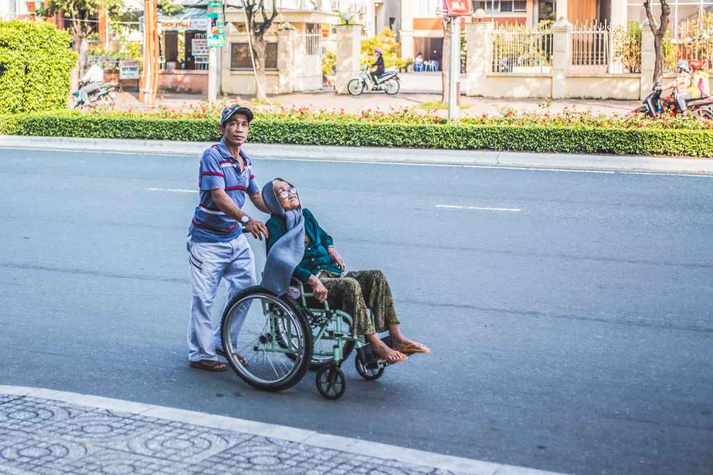
Health Partners and Equipment in Neurologic Rehabilitation Part II

In most cases a neurologic disease or injury occurs suddenly and without warning. The impact can be sharp and wide reaching and few people are prepared for the aftereffects. Recovery and management of such diseases and injuries can be daunting if you do not know what to expect. Many people experience needs or issues that they are unfamiliar with and often are not aware of the various resources and providers available to help them with their needs. Below is a list of resources and community partners you may be able to access where you live to support your recovery and improve your quality of life. By no means is this list exhaustive and I would recommend reaching out to some of the people in your immediate community for more specific guidance. You’d be surprised what resources lie close to home if you know what you are looking for!

If you haven’t checked out Part I of this blog its a great place to start.

Adaptive Equipment

Performing daily tasks such as dressing, walking, meal prep or work tasks may be difficult with a neurologic injury or disorder. Luckily there are creative types out there who have been crafting adaptive devices for almost anything you can think of. A quick internet search may help you find what you are looking for but Occupational, Physical and Speech therapists are also great resources for these devices. Adaptive equipment exists to help with small tasks such as grasping a fork or buttoning a shirt, for bigger tasks such as inserting or removing a urinary catheter, for holding weights during a workout session and many more tasks. 

Splints, bracing and orthotics

Splinting and bracing may also increase your efficiency and independence. Many people view bracing and splinting as a sign that they haven’t recovered or as something that will hinder their recovery but the truth is that when utilized appropriately splinting and bracing may improve your safety, decrease the need for help from others, increase energy, reduce pain and increase ability to walk. Again, physical or occupational therapists are trained in prescribing splints and braces. If you would benefit from a custom brace or splint you may be referred to an Orthotist who will not only work with your insurance to determine if they may pay for some or all of the device but can order an off the shelf brace or build a custom brace to meet your specific needs. A prescription from your physician will be needed if insurance coverage is sought. 

Community Partners and Allied Health Professionals

In addition to your therapy team and medical specialists there are many community partners and allied health professionals that can support you in many ways. Here is just a short list of resources you might find helpful

-Mental/Psychological Support: licensed-clinical social workers, psychologist and psychiatrists can be imperative in supporting mental and emotional wellbeing. Coping and adapting during life with a neurologic injury or disease can be challenging and taking care of your mental and emotional health (that includes caregivers too!) is important. Local support groups are also a great way to find others who share your experience. Everyone of us could use a strong support system and sometimes reaching for support outside of your closest family and friends is important and can help reduce feelings of overwhelm, anxiety or depression.

-Neuropsychologist: Neuropsychologists are different than traditional psychologists. Neuropsychologists study brain and behavior changes that occur following a neurologic trauma or disease. They work specifically to understand the cause and severity of memory difficulties, mood disorders, learning difficulties and nervous system dysfunction. They can assess components such as problem-solving abilities, reasoning, and so forth. They utilize interview, possibly imaging and formalized testing to assess these and many other different areas. Neuropsychological evaluation can give you a deeper understanding of your condition and can help develop a treatment plan to help improve or rehabilitate these impairments. 

-Neuro Ophthalmologist: Neuro Ophthalmologists take care of visual problems related to the nervous system. The brain is highly involved in vision related tasks and any injury or disease that affects the brain or nerves may affect your vision. Neuro Ophthalmology may be beneficial to diagnose visual changes and provide a treatment plan to improve or compensate for your vision impairments.

-Vocational Rehabilitation: Vocational Rehabilitation Programs provide services and support in order to assist persons with disabilities to meet their employment goals. If you have a physical or mental disorder that presents a significant impediment to employment you may qualify for a vocational rehabilitation program. Vocational Rehabilitation Programs are often run or funded by the state and eligibility is determined based on need. An internet search for Vocational Rehabilitation Programs in your state may provide you with the information you need for referral and eligibility to a program. 

Health Partners and Equipment in Neurologic Rehabilitation Part I

In most cases a neurologic disease or injury occurs suddenly and without warning. The impact can be sudden and wide reaching and few people are prepared for the aftereffects. Recovery and management of such diseases and injuries can be daunting if you do not know what to expect. Many people experience needs or issues that they are unfamiliar with and often they are not aware of the various resources and providers available to help them with their needs. Below is a list of resources and community partners you may be able to access where you live to support your recovery and improve your quality of life. By no means is this list exhaustive and I would recommend reaching out to some of the people in your immediate community for more specific guidance. You’d be surprised what resources lie close to home if you know what you are looking for!

Photo by Streetwindy on Pexels.com

During Hospitalization

Nurse case managers and social workers can often be accessed in a hospital or rehabilitation setting to help organize post-acute care and advise you on resources to support you in your community. During hospitalization, decisions about where you or your loved one will be discharged should begin. While getting home is always the ultimate goal sometimes an intermediate level of care is necessary to ensure that return to home is safe and successful for everyone involved.

Post-Acute Care

Several levels of rehabilitation are available to support you in returning to your home and prior level of function. If you can tolerate 3 hours of therapy per day (speech therapy, occupational therapy and/or physical therapy) you may qualify for a stay at an intensive inpatient rehabilitation facility in your area. If you do not meet the qualifications for inpatient rehabilitation or have finished your stay there but are not ready to return home you may be transferred to a skilled nursing facility where you will continue to receive some therapy services and also any continued medical care. Once safe to return home you typically have two options for rehabilitation: home health therapy and outpatient therapies. Which program is right for you depends on many factors but those who can leave the home without difficulty may not qualify for services in the home.

Transportation Services

Several options exist to help transport you to appointments and other important places and likely vary depending on where you live. You may qualify for subsidized transportation through local services like Dial-A-Ride or others like it. Your insurance company may also be able to connect you with a free or low cost ride service based on your needs. Community-based services such as Uber or Lyft may be an option for those that can transfer in and out of a car relatively easily and are safe to travel and navigate this system alone. Reach out to your insurance company and find out what they can offer. Whenever possible, don’t let transportation difficulties stand in the way of getting the care you need.

Mobility Devices

If a mobility device such as a wheelchair, cane or walker is needed before leaving the hospital, often the hospital staff will arrange to have this ordered for you. Wheelchairs may be purchased or rented depending on the need. 

Durable Medical Equipment (DME) companies provide custom wheelchairs, standing frames, gait trainers and other equipment that can assist you in your mobility. Often this type of equipment requires a prescription from a doctor, a formal physical or occupational therapy assessment and formal paperwork to submit to insurance. Depending on your insurance plan they may cover some or all of the cost of this equipment but there are typically limitations on how often large pieces of equipment such as these can be purchased through insurance. If you think you may benefit from a piece of equipment such as this talk to your physician and set up an appointment with a neurologic rehabilitation facility who performs equipment evaluations. 

For smaller devices such as walkers, canes or crutches individuals are often responsible for purchasing these on their own. There are many options out there, however, and if you are unsure which one is best for you talk with your therapist. They may have a variety of assistive devices at their clinic that you can try and they can advise you on what might be best for you at a particular stage of your recovery.  

Return to Driving

Determining when it is safe and appropriate to return to driving is a question that many people face. Driving is a highly skilled activity that requires high level physical and cognitive abilities and has the potential to injure the drivers and others around him/her if undertaken inappropriately. There may be several resources in your area that can assist in determining readiness to drive. The first step is to speak with your physician and gain clearance from them. You may also need to reach out to your local Department of Motor Vehicles for re-licensing or testing. Local Occupational Therapist facilities may have formal return-to-driving assessment programs that can assess various aspects of driving or there may be local companies who specialize in driving assessment and training after injury or illness.

Another consideration is adaptation to the traditional driving mechanisms. Most cars can be adapted with hand controls or other modifications for those that cannot use their hands or legs to drive in the traditional way. Wheelchair accessible vans are also an option for wheelchair users and may be fitted with ramps or options to drive from the wheelchair itself. Search the internet for adaptive driving companies locally or ask your physical or occupational therapist for a recommendation. 

Home Modifications

Oftentimes after a neurologic injury or event individuals may require adaptive equipment and home modifications. A great first step is to ask your rehabilitation team to recommend equipment that may be beneficial for you and your family. Local medical supply stores and even amazon.com are great resources for equipment. You may need to find a handy friend or a licensed contractor to make bigger installations such as ramps, stair lifts or grab rails.  

As you can see, there are many resources out there to assist you in returning to your home and your day to day life. Check out Part II of this Blog to see even more ideas.